At a glance
Matrix-assisted laser desorption ionization time of flight (MALDI-TOF) is one of the easiest and most accurate methodologies for identifying yeasts. This SOP outlines the steps for identification of yeasts. It also includes instructions on how to access an expanded database in MicrobeNet.
1.0 Purpose
This document outlines the procedure for identification of yeast isolates using the MALDI-ToF MS Bruker Biotyper platform and a custom extraction protocol optimized to identify isolates that are represented in the Bruker database and the MicrobeNet database.
2.0 Definitions
- MALDI-ToF: Matrix-assisted laser ionization (time of flight)
- SAB Media: Sabouraud's Dextrose agar
- Chromagar Media: CHROMagar Candida Chromogenic agar
- BTS: Bacterial Test Standard
4.0 Reagents, supplies, and media
4.1 100% Ethanol-Sterile Solution: example, SIGMA (Catalog # 459836), or equivalent. Always use Ethanol labeled "molecular biology reagent."
4.2 Sterile WFI Quality Cell Culture Grade Water: example, Fisher Scientific (Product # L4321849), or equivalent
4.3 Sabouraud's Dextrose Agar (Emmons): Plates and slants; example, Fisher Scientific (Product #L21827), or equivalent
4.4 CHROMagar Candida Chromogenic agar: available from multiple vendors as plates or powder
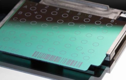
4.5 Reusable polished steel MALDI target plate with 96 sample positions (Bruker Product # 8280800)
4.6 Bruker matrix HCCA (HCCA = α-Cyano-4-hydroxycinnamic acid): (Product # 255344) — Prepare by adding 250 µl of Bruker Standard Solvent. Vortex until liquid is clear.
4.7 70% Formic Acid: example, SIGMA (Catalogue # 27001), diluted using sterile WFI Quality cell culture grade water
4.8 Bacterial test standard (BTS): Bruker (Product # 255343) — Prepare BTS according to manufacturer's instructions and store at ≤ -18°C until ready for use.
4.9 Bruker Standard Solvent (Sigma Aldrich #900666)
4.10 1µl Culture loops
4.11 1.5 mL microcentrifuge tubes
5.0 Safety
NOTE: All institutional safety procedures must be followed in the performance of this standard operating procedure.
5.1 Standard personal protective equipment should follow institutional guidelines but should consist of at least gowns, gloves, and safety glasses, which should be worn at all times.
5.2 All of the set-up procedures should be performed in a biological safety cabinet.
5.3 Chemical Safety
- 5.3.1 Formic Acid: Flammable, Toxic, and Corrosive
- 5.3.2 α-Cyano-4-hydroxycinnamic acid (HCCA): Skin irritant, serious eye irritant
6.0 Sample preparation
6.1 Growth
- 6.1.1 Overnight growth of a pure yeast culture should be used for routine microorganism identification.
- 6.1.2 Slow-growing yeast (e.g., Candida haemulonii) may need to grow for several days before testing.
6.2 Storage
- 6.2.1 Store plates at room temperature for 1-5 days is acceptable.
- 6.2.2 Culture medium (Sabouraud dextrose agar or CHROMagar) and growth temperature (25°C vs 37°C) have little to no effect on results.
NOTE: Do not use organisms that have been stored at 4°C or lower as this has a negative impact on quality of spectra and reproducibility.
7.0 Quality control
7.1 For each run, include pure culture of Candida auris and Candida parapsilosis, in duplicate, as positive controls.
8.0 Procedure
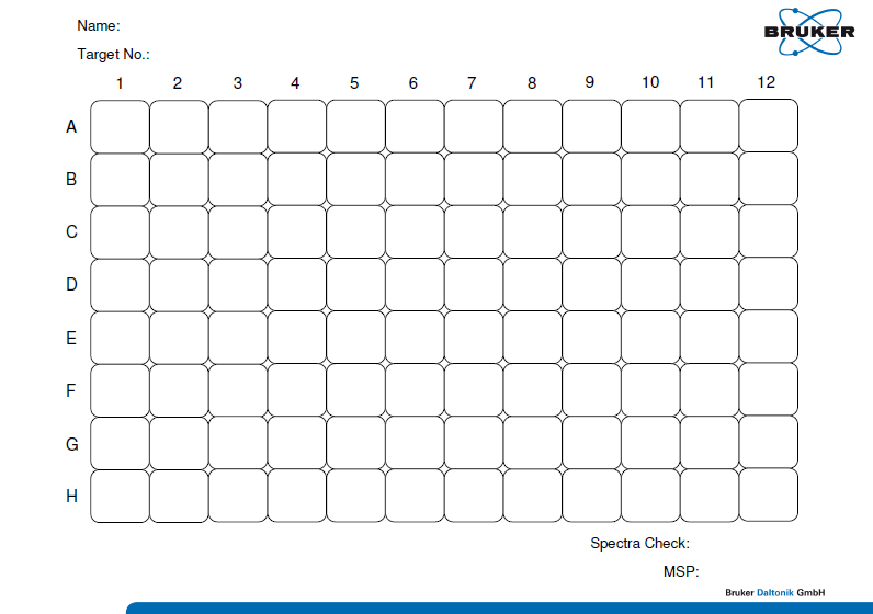
NOTE: Prior to beginning the procedure, note on a sample key spreadsheet (Appendix) the location of where each specimen will be placed on the 96-spot reusable stainless steel target plate.
8.1 Extraction procedure
NOTE: Perform the following steps in a biological safety cabinet to ensure safety and sterility.
- 8.1.1 Perform extraction procedure in duplicate.
- 8.1.2 Label two 1.5 mL microcentrifuge tubes for each sample, including QC strains from Step 6.1.
- 8.1.3 Add 50 µL of sterile WFI quality water to each tube.
- 8.1.4 Using a sterile, clear 1 µl inoculating loop, transfer 1 loopful of microorganism to both tubes containing 50 µl of sterile water. If the colonies are small, transfer more than one colony, choosing isolated colonies, when possible.
- 8.1.5 Secure the lid of the tube.
- 8.1.6 Add 50 μL of 100% Ethanol to each tube.
- 8.1.7 Using a 100 µl pipette set to 50 µl, pipette the liquid up and down 10 times to homogenize.
- 8.1.8 From each tube, add one microliter of the resulting suspension to a 96-spot reusable stainless steel target plate (see Fig. 1).
- 8.1.9 Let the plate air dry for two minutes.
- 8.1.10 Overlay dried spot with 1 μL of 70% Formic Acid.
- 8.1.11 Air dry for two minutes.
- 8.1.12 Add 1 μL of the BTS suspension to two open spots on the 96-spot reusable stainless steel target plate (do not add formic acid to BTS! Simply allow to air dry and proceed to step 8.1.13). Perform this step once a week. For preparation of BTS, see step 4.8.
- 8.1.13 As soon as spots are dry, immediately overlay with 1 μL of HCCA matrix. Do not delay the addition of HCCA matrix as it will decrease the quality of your run.
- 8.1.14 Air dry for two minutes.
- 8.1.15 As soon as spots are dry, they are ready to be subject to MALDI-TOF analysis. Target must be completely dry before it is inserted into MALDI BioTyper.
- 8.1.16 Once the target plate is dry, the plate must be run within 24 hours.
- 8.1.17 Be sure that the location of each specimen on the 96-spot reusable stainless steel target plate is recorded on a sample key spreadsheet. Verify the spreadsheet numbers to make sure they match the original sample numbers.
- 8.1.18 Samples are now ready for the MALDI-ToF MS assay on the Bruker Biotyper platform.
- 8.1.19 Resulting spectra must be interpreted using Bruker database.
9.0 Performance specifications
9.1 Bruker allows for any score above 2.0 to be acceptable for a species identification. A laboratory may perform an in-house validation to determine a lower score which may be acceptable for species identification.

10.0 Interpreting results
10.1 Review control strains.
- 10.1.1 At least one of the duplicates for each control strain must generate a score of ≥ 2 with a correct identification (no conflicting results between the duplicates).
- 10.1.2 If the control strains generate scores < 2 or incorrect identification, the results cannot be interpreted, and the extraction/submission process must be repeated.
10.2 Candida species will be identified if database match scores are ≥ 2 for one or more of the duplicates.
10.3 If no species identification is made, repeat extraction/submission process using a new subculture.
10.4 If no identification is successful after second submission, the isolate must be identified by another methodology.
(Optional) 11.0 Submitting main spectra to MicrobeNet
11.1 Go to the MicrobeNet website.
11.2 Request a MicrobeNet account.
11.3 Log into account.
11.4 Convert Main Spectra to XML file on your Bruker software.
11.5 Drag and drop the XML file onto the MicrobeNet website.
11.6 The top 10 matches will populate.
Disclaimer
The Mycotic Diseases Laboratory developed this document as an example test procedure for identifying yeast isolates using MALDI-TOF. It is the responsibility of the testing laboratory to ensure content and format are modified as necessary to meet applicable regulatory requirements, quality management system standards, and chemical, radiological and biological safety requirements. This is not a controlled document, and the described test methods are subject to change without notice. It is the responsibility of the testing laboratory to ensure the information within this document remains applicable. Contact the test developer at [email protected] to find out whether any changes have been made.
Use of trade names and commercial sources are for identification only and do not constitute endorsement by the Public Health Service, the United States Department of Health and Human Services, or the Centers for Disease Control and Prevention.

